
Education, Research & Development
Bone Case 3
Details on the Request Card
53 year old man with non-small cell carcinoma with previous carcinoma. Recent CT showed liver and adrenal metastases. Low right posterior chest wall pain. ?rib metastases.Procedure
Anterior and posterior images are shown, 3hrs after injection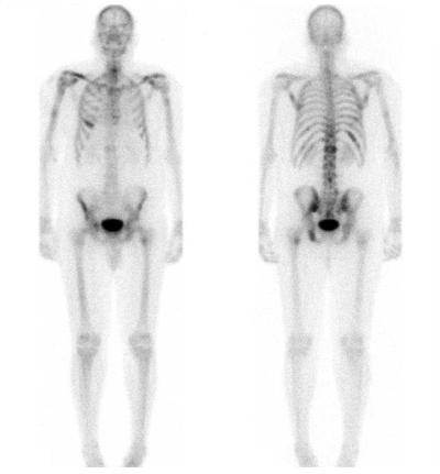
Questions
1) Are there any abnormal areas of uptake?
2) If so, where?
3) What are the advantages of this technique over CT?
The text is entirely the opinion of the author and does not necessarily reflect that of RUH NHS Trust or the Bristol Radiology Training Scheme. Website content devised by Paul McCoubrie.